Abstract
Research Article
In silico analysis and characterization of fresh water fish ATPases and homology modelling
Rumpi Ghosh, AD Upadhayay and AK Roy*
Published: 11 October, 2017 | Volume 1 - Issue 1 | Pages: 018-024
ATPases is known to be a crucial in many biological activities of organisms. In this study, physicochemical properties and modeling of ATPases protein of fish was analysed using In silico approach. ATPases a protein selected from fish species, including Gold fish (Carassius auratus auratus), Zebra fish (Hypancistrus zebra), White fishes (Coregonus autumnalis), Grass carp (Ctenopharyngodon idella) and Anabas testudineus (Koi) were used in this study. Physicochemical characteristics showed with molecular weight (25045.58-25148.57Da), theoretical isoelectric point (9.30-9.97), extinction coefficient(26470-34950), aliphatic index(147.31-150.35), instability index(32.84-42.67), total number of negatively charged residues and positively charged residues (5/7-6/8), and grand average of hydropathicity (1.014-1.151) were computed. All proteins were classified as transmembrane proteins. In secondary structure prediction, all proteins were composed of random coils as predominant, followed by extended strands, alpha helix and beta turn. Three dimensional structure of protein were predicted and verified as good structures. All model structures were evaluated being accepted and reliable based on structural evaluation and stereo chemical analysis.
Read Full Article HTML DOI: 10.29328/journal.hpbr.1001003 Cite this Article Read Full Article PDF
Keywords:
ATPase; Expasy’s prot; Physicochemical characterisation; Clustal W; Modelling etc
References
- Tran Ngoc Tuan, Pham Minh Duc. In silico analysis of freshwater fish major histocompatibility complex Class II Alpha. Asia pacific journal of science and technology. 2016; 21: 1-8. Ref.: https://goo.gl/MtFwo3
- Tran Ngoc Tuan, Wang Gui-Tang, Pham Minh duc. Tumor necrosis factor alpha of teleosts: In silico characterization and homology modeling. Songklanakarin Journal of Science & Technology. 2016; 38: 549-557. Ref.: https://goo.gl/YB3dgD
- Tamura K, Stecher G, Peterson D, Filipski A, Kumar S. MEGA6: Molecular evolutionary genetics analysis version 6.0. Molecular biology and Evolution. 2013; 30: 2725-2729. Ref.: https://goo.gl/PYMja7
- Hogg PJ. Disulfide bonds as switches for protein function. Trends Biochem Sci. 2003; 28: 210-214. Ref.: https://goo.gl/5EevyN
- Geourjon C, Deleage G. SOPMA: significant improvements in protein secondary structure prediction by consensus prediction from multiple alignments. Computer application in bioscience. 1995; 11: 681-684. Ref.: https://goo.gl/wUsYbn
- Schwede T, Kopp J, Guex N, Peitsch MC. Swiss-MODEL:An automated protein homology modeling server. Nucleic acids research. 2003; 31: 3381-3385. Ref.: https://goo.gl/MjY825
- Arnold K, Bordoli L, Kopp J, Schwede T. The Swiss-Model workspace: A web-based environment for protein structure homology modelling. Bioinformatics. 2006; 22: 195-201. Ref.: https://goo.gl/tTxBtk
- Fiser A. Template-based Protein structure modeling. In computational Biology. 2010; 73-94.
- Template-based protein structure modeling. D Fenyo. Computational Biology: Humana press; 2004.
- Ramachandran GN, Ramakrisnan C, Sasisekhran V. Stereochemistry of polypeptide chain configurations. J Mol Biol. 1963; 7: 95-99. Ref.: https://goo.gl/BaHh8q
- Laskowski RA, Rullman JAC, MacArthur MW, Kaptein R, Thornton JM. AQUA and PROCHECK_NMR: Programs for checking the quality of protein structures solved by NMR. Journal of bimolecular NMR. 1996; 8: 477-486. Ref.: https://goo.gl/7Emfju
- Ramachandran G, Ramakrishnan c, Sasisekharan V. Stereochemistry of polypeptide chain configurations. J Mol Biol. 1963; 7: 95-99. Ref.: https://goo.gl/W7vzZ9
- Wallner B, Elofsson A. Can correct protein models be identified? Protein science. 2003; 12: 1073-1086. Ref.: https://goo.gl/VrWtw2
- Wiederstein M, Sippl MJ. ProSA-web: Interactive web service for the recognition of errors in three dimensional structures of proteins. Nucleic Acids Res. 2007; 35: W407-W410. Ref.: https://goo.gl/QFtmm8
- Sippl MJ. Recognition of errors in three dimensional structures of proteins. Proteins: structure, function and genetics. 1993; 17: 355-362. Ref.: https://goo.gl/cqfoDW
- Yadav NK, Shukla P, Parrek S, Singh R. Structure prediction and functional characterization of matrix protein of human Metapneumovirus (Strain can97-83)(hMPV). International Journal of advanced Life sciences. 2013; 6: 538-548. https://goo.gl/GDGdsz
- Ghosh R, Upadhayay AD, Roy AK. In silico Analysis, structure modelling and Phosphorylation site prediction of Vitellogenin Protein from Gibelion catla. Journal of applied Biotechnology and bioengineering. 2017; 3: 55.
- Pradeep N, Anupama A,Vidyashree K, Lakshmi P. In silico characterization of industrial important cellulases using computational tools. Advances science and Tecnology. 2012; 48: 14.
- Rumpi G, Upadhyay AD, Roy AK, Samik A. Structural analysis of cytochrome C Genes of Major Carp and utility of Forensic Investigation. Journal of Forensic and crime investigation. 2017; 1: 103. Ref.: https://goo.gl/ALvudA
- Tuan TN, WeiMin W, Duc PM. Characterization and homology modeling of finfish NK-kappa B inhibitor alpha using In silico analysis. J Sci Develop. 2015; 13: 216-225. Ref.: https://goo.gl/gqRHkP
- Ghosh R, Upadhayay AD, Roy AK, Mamta singh. In silico Analysis and 3d Structure Prediction of mitochondrial RHO GTPase 2 Protein of Danio rerio (zebra fish) by Homology Modeling. Jacobs J Bioinform Proteom. 2016; 1: 006. Ref.: https://goo.gl/VPmbwg
- Ghosh R, Upadhayay AD, Roy AK. In silico analysis of Rag 1 Protein of Labeo calbasu. International Journal of Innovative Research in Science & Engineering. 4: 136. Ref.: https://goo.gl/Wmo4w8
Figures:
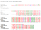
Figure 1

Figure 2

Figure 3
Similar Articles
-
In silico analysis and characterization of fresh water fish ATPases and homology modellingRumpi Ghosh,AD Upadhayay,AK Roy*. In silico analysis and characterization of fresh water fish ATPases and homology modelling. . 2017 doi: 10.29328/journal.hpbr.1001003; 1: 018-024
Recently Viewed
-
Metastatic Brain Melanoma: A Rare Case with Review of LiteratureNeha Singh,Gaurav Raj,Akshay Kumar,Deepak Kumar Singh,Shivansh Dixit,Kaustubh Gupta*. Metastatic Brain Melanoma: A Rare Case with Review of Literature. J Radiol Oncol. 2025: doi: 10.29328/journal.jro.1001080; 9: 050-053
-
Validation of Prognostic Scores for Attempted Vaginal Delivery in Scar UterusMouiman Soukaina*,Mourran Oumaima,Etber Amina,Zeraidi Najia,Slaoui Aziz,Baydada Aziz. Validation of Prognostic Scores for Attempted Vaginal Delivery in Scar Uterus. Clin J Obstet Gynecol. 2025: doi: 10.29328/journal.cjog.1001185; 8: 023-029
-
Scientific Analysis of Eucharistic Miracles: Importance of a Standardization in EvaluationKelly Kearse*,Frank Ligaj. Scientific Analysis of Eucharistic Miracles: Importance of a Standardization in Evaluation. J Forensic Sci Res. 2024: doi: 10.29328/journal.jfsr.1001068; 8: 078-088
-
A study of coagulation profile in patients with cancer in a tertiary care hospitalGaurav Khichariya,Manjula K*,Subhashish Das,Kalyani R. A study of coagulation profile in patients with cancer in a tertiary care hospital. J Hematol Clin Res. 2021: doi: 10.29328/journal.jhcr.1001015; 5: 001-003
-
Additional Gold Recovery from Tailing Waste By Ion Exchange ResinsAshrapov UT*, Malikov Sh R, Erdanov MN, Mirzaev BB. Additional Gold Recovery from Tailing Waste By Ion Exchange Resins. Int J Phys Res Appl. 2024: doi: 10.29328/journal.ijpra.1001098; 7: 132-138
Most Viewed
-
Evaluation of Biostimulants Based on Recovered Protein Hydrolysates from Animal By-products as Plant Growth EnhancersH Pérez-Aguilar*, M Lacruz-Asaro, F Arán-Ais. Evaluation of Biostimulants Based on Recovered Protein Hydrolysates from Animal By-products as Plant Growth Enhancers. J Plant Sci Phytopathol. 2023 doi: 10.29328/journal.jpsp.1001104; 7: 042-047
-
Sinonasal Myxoma Extending into the Orbit in a 4-Year Old: A Case PresentationJulian A Purrinos*, Ramzi Younis. Sinonasal Myxoma Extending into the Orbit in a 4-Year Old: A Case Presentation. Arch Case Rep. 2024 doi: 10.29328/journal.acr.1001099; 8: 075-077
-
Feasibility study of magnetic sensing for detecting single-neuron action potentialsDenis Tonini,Kai Wu,Renata Saha,Jian-Ping Wang*. Feasibility study of magnetic sensing for detecting single-neuron action potentials. Ann Biomed Sci Eng. 2022 doi: 10.29328/journal.abse.1001018; 6: 019-029
-
Pediatric Dysgerminoma: Unveiling a Rare Ovarian TumorFaten Limaiem*, Khalil Saffar, Ahmed Halouani. Pediatric Dysgerminoma: Unveiling a Rare Ovarian Tumor. Arch Case Rep. 2024 doi: 10.29328/journal.acr.1001087; 8: 010-013
-
Physical activity can change the physiological and psychological circumstances during COVID-19 pandemic: A narrative reviewKhashayar Maroufi*. Physical activity can change the physiological and psychological circumstances during COVID-19 pandemic: A narrative review. J Sports Med Ther. 2021 doi: 10.29328/journal.jsmt.1001051; 6: 001-007

HSPI: We're glad you're here. Please click "create a new Query" if you are a new visitor to our website and need further information from us.
If you are already a member of our network and need to keep track of any developments regarding a question you have already submitted, click "take me to my Query."


